Document Type : Original Article
Authors
1 Department of Biological Science Technology, Federal Polytechnic Mubi, PMB 035 Mubi, Adamawa State, Nigeria
2 Department of Microbiology, Adamawa State University, Mubi, Adamawa State, Nigeria
3 Department of Biomedical and Pharmaceutical Technology, Federal Polytechnic Mubi, Adamawa State, Nigeria
Abstract
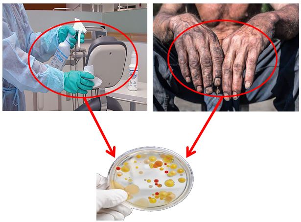
The ability of the bacterial isolate to cause debilitating effects on the host is intricate and is a function of many factors, particularly that of the host and the bacteria. Among the bacterial factors are the virulence mechanisms. As such this research was a cross-sectional study conducted between October–December 2021 to establish the existence of virulence determinants on bacterial isolates from hospital fomites and the hands of healthcare workers. To achieve this, 100 samples (including sink, beddings, door handles, benches, and hands of healthcare workers) from children, female and male wards of Mubi General Hospital were analyzed for bacterial growth and were identified by standard procedure. Isolates were subsequently screened for virulent determinants (hemolysis, hemagglutination, biofilm production, and heteroresistance) phenotypically by standard methods. From the 72 bacterial isolates recovered, 23(31.9%) were biofilm-producing organisms. Of these, 15(20.8%) and 8(11.1%) were moderate and high biofilm-producing organisms respectively with no statistical difference (P=0.665). Pseudomonas aeruginosa (13.9%) was the most predominant biofilm-producing organism. Furthermore, hemolysin production was predominant in Staphylococcus aureus (71.4%), while positive hemagglutination reaction was predominant in P. aeruginosa (38.5%). Sixteen (16) bacterial isolates showed heteroresistance (HR) to various antibiotics; of these, Escherichia coli (43.8%) constitute the majority of the isolates. The expression of such virulence determinants by bacterial isolates in the study area may constitute a health risk to patients and hamper the quality of health care delivery.
Graphical Abstract
Keywords
Main Subjects
Selected author of this article by journal
.jpg)
Federal Polytechnic Mubi
Google Scholar
Open Access
This article is licensed under a CC BY License, which permits use, sharing, adaptation, distribution and reproduction in any medium or format, as long as you give appropriate credit to the original author(s) and the source, provide a link to the Creative Commons license, and indicate if changes were made. The images or other third party material in this article are included in the article’s Creative Commons license, unless indicated otherwise in a credit line to the material. If material is not included in the article’s Creative Commons license and your intended use is not permitted by statutory regulation or exceeds the permitted use, you will need to obtain permission directly from the copyright holder. To view a copy of this license, visit: http://creativecommons.org/licenses/by/4.0/
Publisher’s Note
CMBR journal remains neutral with regard to jurisdictional claims in published maps and institutional afflictions.
Letters to Editor
Given that CMBR Journal's policy in accepting articles will be strict and will do its best to ensure that in addition to having the highest quality published articles, the published articles should have the least similarity (maximum 15%). Also, all the figures and tables in the article must be original and the copyright permission of images must be prepared by authors. However, some articles may have flaws and have passed the journal filter, which dear authors may find fault with. Therefore, the editor of the journal asks the authors, if they see an error in the published articles of the journal, to email the article information along with the documents to the journal office.
CMBR Journal welcomes letters to the editor ([email protected], [email protected]) for the post-publication discussions and corrections which allows debate post publication on its site, through the Letters to Editor. Critical letters can be sent to the journal editor as soon as the article is online. Following points are to be considering before sending the letters (comments) to the editor.
[1] Letters that include statements of statistics, facts, research, or theories should include appropriate references, although more than three are discouraged.
[2] Letters that are personal attacks on an author rather than thoughtful criticism of the author’s ideas will not be considered for publication.
[3] There is no limit to the number of words in a letter.
[4] Letter writers should include a statement at the beginning of the letter stating that it is being submitted either for publication or not.
[5] Anonymous letters will not be considered.
[6] Letter writers must include Name, Email Address, Affiliation, mobile phone number, and Comments.
[7] Letters will be answered as soon as possible.
- Ige O, Jimoh O, Ige S, Ijei I, Zubairu H, Olayinka A (2021) Profile of bacterial pathogens contaminating hands of healthcare workers during daily routine care of patients at a tertiary hospital in northern Nigeria. African Journal of Clinical and Experimental Microbiology 22 (1): 103-108. doi:https://dx.doi.org/10.4314/ajcem.v22i1.14
- Nguyen VV, Dong HT, Senapin S, Pirarat N, Rodkhum C (2016) Francisella noatunensis subsp. orientalis, an emerging bacterial pathogen affecting cultured red tilapia (Oreochromis sp.) in Thailand. Aquaculture Research 47 (11): 3697-3702. doi:https://doi.org/10.1128/CMR.00059-12
- Sharma AK, Dhasmana N, Dubey N, Kumar N, Gangwal A, Gupta M, Singh Y (2017) Bacterial virulence factors: secreted for survival. Indian journal of microbiology 57 (1): 1-10. doi:https://doi.org/10.1007/s12088.016-0625-1
- Cepas V, Soto SM (2020) Relationship between virulence and resistance among gram-negative bacteria. Antibiotics 9 (10): 719. doi:https://doi.org/10.3390/antibiotics9100719
- Leitão JH (2020) Microbial virulence factors. Int J Mol Sci 21 (15): 5320. doi:https://doi.org/10.3390/ijms21155320
- Tula MY, Enabulele OI, Ophori AE, Aziegbemhin AS, Iyoha O, Filgona J (2022) Phenotypic and molecular detection of multi-drug resistant Enterobacteriaceae species from water sources in Adamawa-North senatorial zone, Nigeria. DYSONA-Life Science 3 (2): 57-68. doi:https://doi.org/10.30493/DLS.2022.351097.
- Tula MY, Onyeje GA, John A (2018) Prevalence of Antibiotic Resistant and Biofilm Producing Escherichia coli and Salmonella spp from Two Sources of Water in Mubi, Nigeria. Frontier in Science 8 (1): 18-25. doi:https://doi.org/10.5923/j.fs.20180801.03
- Clinical, Institute LS (2017) Performance standards for antimicrobial susceptibility testing. Clinical and Laboratory Standards Institute Wayne, PA,
- El-Halfawy OM, Valvano MA (2015) Antimicrobial heteroresistance: an emerging field in need of clarity. Clinical microbiology reviews 28 (1): 191-207. doi:https://doi.org/10.1128/CMR.00058-14
- Valliammai A, Sethupathy S, Ananthi S, Priya A, Selvaraj A, Nivetha V, Aravindraja C, Mahalingam S, Pandian SK (2020) Proteomic profiling unveils citral modulating expression of IsaA, CodY and SaeS to inhibit biofilm and virulence in Methicillin-resistant Staphylococcus aureus. International Journal of Biological Macromolecules 158): 208-221. doi:https://doi.org/10.1016/j.ijbiomac.2020.04.231
- Vagarali MA, Karadesai SG, Patil CS, Metgud SC, Mutnal MB (2008) Haemagglutination and siderophore production as the urovirulence markers of uropathogenic Escherichia coli. Indian Journal of Medical Microbiology 26 (1): 68-70. doi:https://doi.org/10.1016/S0255-0857(21)01997-6
- Hassan R, El-Naggar W, El-Sawy E, El-Mahdy A (2011) Characterization of some virulence factors associated with Enterbacteriaceae isolated from urinary tract infections in Mansoura Hospitals. Egypt J Med Microbiol 20 (2): 9-17
- Rajkumar H, Devaki R, Kandi V (2016) Comparison of hemagglutination and hemolytic activity of various bacterial clinical isolates against different human blood groups. Cureus 8 (2). doi:https://doi.org/10.7759/cureus.489
- Persson G, Bojesen AM (2015) Bacterial determinants of importance in the virulence of Gallibacterium anatis in poultry. Veterinary Research 46 (1): 1-11. doi:https://doi.org/10.1186/s13567-015-0206-z
- Chung PY (2016) The emerging problems of Klebsiella pneumoniae infections: carbapenem resistance and biofilm formation. FEMS microbiology letters 363 (20): fnw219. doi:https://doi.org/10.1093/femsie/fnw219
- Murphy CN, Clegg S (2012) Klebsiella pneumoniae and type 3 fimbriae: nosocomial infection, regulation and biofilm formation. Future Microbiology 7 (10): 1234-1234. doi:https://doi.org/10.2217/fmb.12.74.
- Hogan JS, Todhunter DA, Smith KL, Schoenberger PS (1990) Hemagglutination and Hemolysis by Escherichia coli Isolated from Bovine Intramammary Infections1. Journal of Dairy Science 73 (11): 3126-3131. doi:https://doi.org/10.3168/jds.S0022-0302(90)79001-7
- Swarna S, Gomathi S (2017) Biofilm Production in Carbapenem Resistant Isolates from Chronic Wound Infections. International Journal of Medical Research & Health Sciences 6 (2): 61-67
- Dumaru R, Baral R, Shrestha LB (2019) Study of biofilm formation and antibiotic resistance pattern of gram-negative Bacilli among the clinical isolates at BPKIHS, Dharan. BMC Research Notes 12 (1): 1-6. doi:https://doi.org/10.1186/s13104-019-4084-8
- Allam N (2017) Correlation between Biofilm Production and Bacterial UrinaryTract Infections: New Therapeutic Approach. Egyptian Journal of Microbiology 52 (1): 39-48. doi:https://doi.org/10.21608/EJM.2017.1014.1021
- Shrestha LB, Bhattarai NR, Khanal B (2018) Comparative evaluation of methods for the detection of biofilm formation in coagulase-negative staphylococci and correlation with antibiogram. Infection and drug resistance 11): 607. doi:https://doi.org/10.2147/IDR.S159764
- SARKAR S, DUTTA S, NAMHATA A, BANERJEE C, SENGUPTA M, SENGUPTA M (2020) Beta-lactamase Profile and Biofilm Production of Pseudomonas aeruginosa Isolated from a Tertiary Care Hospital in Kolkata, India. Journal of Clinical & Diagnostic Research 14 (10): 22-27. doi:https://doi.org/10.7860/JCDR/2020/44128.14161
- Kukhtyn M, Berhilevych O, Kravcheniuk K, Shynkaruk O, Horyuk Y, Semaniuk N (2017) The influence of disinfectants on microbial biofilms of dairy equipment. Antimicrobial agents and chemotherapy 45 (4): 999-1007. doi:https://doi.org/10.1128/AAC.45.4.999-1007.2001
- Macia MD, Rojo-Molinero E, Oliver A (2014) Antimicrobial susceptibility testing in biofilm-growing bacteria. Clinical Microbiology and Infection 20 (10): 981-990. doi:https://doi.org/10.1111/1469-0691.12651
- Andersson DI, Nicoloff H, Hjort K (2019) Mechanisms and clinical relevance of bacterial heteroresistance. Nature Reviews Microbiology 17 (8): 479-496. doi:https://doi.org/10.1038/s41579-019-0218-1.
- Band VI, Weiss DS (2021) Heteroresistance to beta-lactam antibiotics may often be a stage in the progression to antibiotic resistance. PLoS Biology 19 (7): e3001346. doi:https://doi.org/10.1371/journal.pbio.3001346
- Brauner A, Fridman O, Gefen O, Balaban NQ (2016) Distinguishing between resistance, tolerance and persistence to antibiotic treatment. Nature Reviews Microbiology 14 (5): 320-330. doi:https://doi.org/10.1038/nrmicro.2016.34
- Falagas ME, Makris GC, Dimopoulos G, Matthaiou DK (2008) Heteroresistance: a concern of increasing clinical significance? Clinical Microbiology and Infection 14 (2): 101-104. doi:https://doi.org/10.1111/j.1469-0691.2007.01912.x
- Mesev EV, LeDesma RA, Ploss A (2019) Decoding type I and III interferon signalling during viral infection. Nature microbiology 4 (6): 914-924. doi:https://doi.org/10.1038/s41564-019-0480-z


